
Rubidium is a very reactive element that was first discovered in 1861. It has many uses from serving as a photocell (a type of detector that senses light) to being used as a frequency standard in atomic clocks. In more recent years, rubidium has been used in the medical field for diagnostic testing of coronary heart disease.

Figure 1: Illustration of a healthy artery compared to an artery with plaque build-up. Taken from reference (2).
Coronary artery disease (CAD) is the most common type of heart disease and is a leading cause of death globally.1 In the United States alone, 13 million people are affected by CAD. This disease develops when deposits of cholesterol (plaque) builds up on the inner walls of the coronary arteries, types of arteries that supply blood to the heart (Fig 1.) Plaque build-up narrows and hardens the coronary arteries which decreases blood flow and oxygen transport to the heart. Over time, CAD can weaken the heart, leading to shortness of breath, angina, or heart failure. Coronary artery disease develops over decades, meaning it can go unnoticed until severe symptoms arise. Consequently, the need for effective and reliable detection and early diagnostic testing is immense.
A new technique that combines the imaging technology of PET (positron emission tomography) and CT (computerized tomography) scans that use rubidium has shown promising results for increased accuracy and sensitivity for detecting CAD.3,4
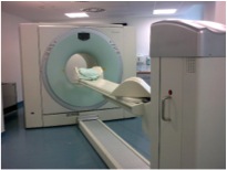
Figure 2: Image of an integrated PET-CT scanner. Taken from reference (2.)
PET scans work by using a small amount of a radioactive substance, called a tracer, to create images of the body.5 Rubidium can be used a radioactive tracer because it has a similar structure to potassium, which is naturally absorbed by body tissue. Furthermore, rubidium decays quickly, meaning it remains in the body for only a short period of time. This is desirable because having radioactive substances in the body’s system for a prolonged time could have negative side effects. To form the PET image the tracer, rubidium, is injected into the body and then is given time to be absorbed by organs and tissues of interest.5 The image is then constructed by looking at different concentrations of the absorbed rubidium in the organ or tissue. PET scans are particularly useful because they can show where there is poor blood flow to the heart.
CT is an imaging method that uses x-rays to form pictures of the body.5 CT images are processed by a computer to create cross-section views of parts of the body, such as the heart. PET and CT scans can be integrated so that they acquire images sequentially and get superimposed into the same image. Studies have shown that integrated PET-CT scans that use rubidium produce very sensitive and accurate images that can be used to diagnose CAD.3,4 In fact, PET-CT scans using rubidium can be more sensitive and accurate than other types of diagnostic testing that have been used for detecting CAD in previous years.3,6 As such, rubidium PET-CT scans have become increasingly useful and well-accepted in the medical community. Combining PET and CT technology allows medical professionals to make better-informed diagnostic decision because the integrated PET-CT image provides more information than individual images could provide.
References:
(1) Mayo Clinic. http://www.mayoclinic.org/
(2) Wikipedia Commons. https://commons.wikimedia.org/wiki
(3) Sampson, U.K.; Dorbala, S.; Limaye, A.; Kwong, R.; Di Carli, M.F. J. Am. Coll. Cardiol. 2007, 49, 1052-58.
(4) Nakazato, R.; Berman, D.; Dey, D.; Meunier, L.; Hayes, S.; Fermin, J.; Cheng, V.; Thomson, L.; Friedman, J.; Germano, G.; Slomka, P. J. Nuc. Cardiol. 2012, 2, 265-276.
(5) Medline Plus: National Institutes of Health. http://www.nlm.nih.gov/
(6) Go, R.T.; Marwick, T.H.; MacIntyre, W.J. Saha, G.B.; Neumann, D.R.; Underwood, D.A.; Simpfendorfer, C.C. J. Nucl. Med. 1990, 31, 1899-1905.